How are spect and pet used in medical imaging?
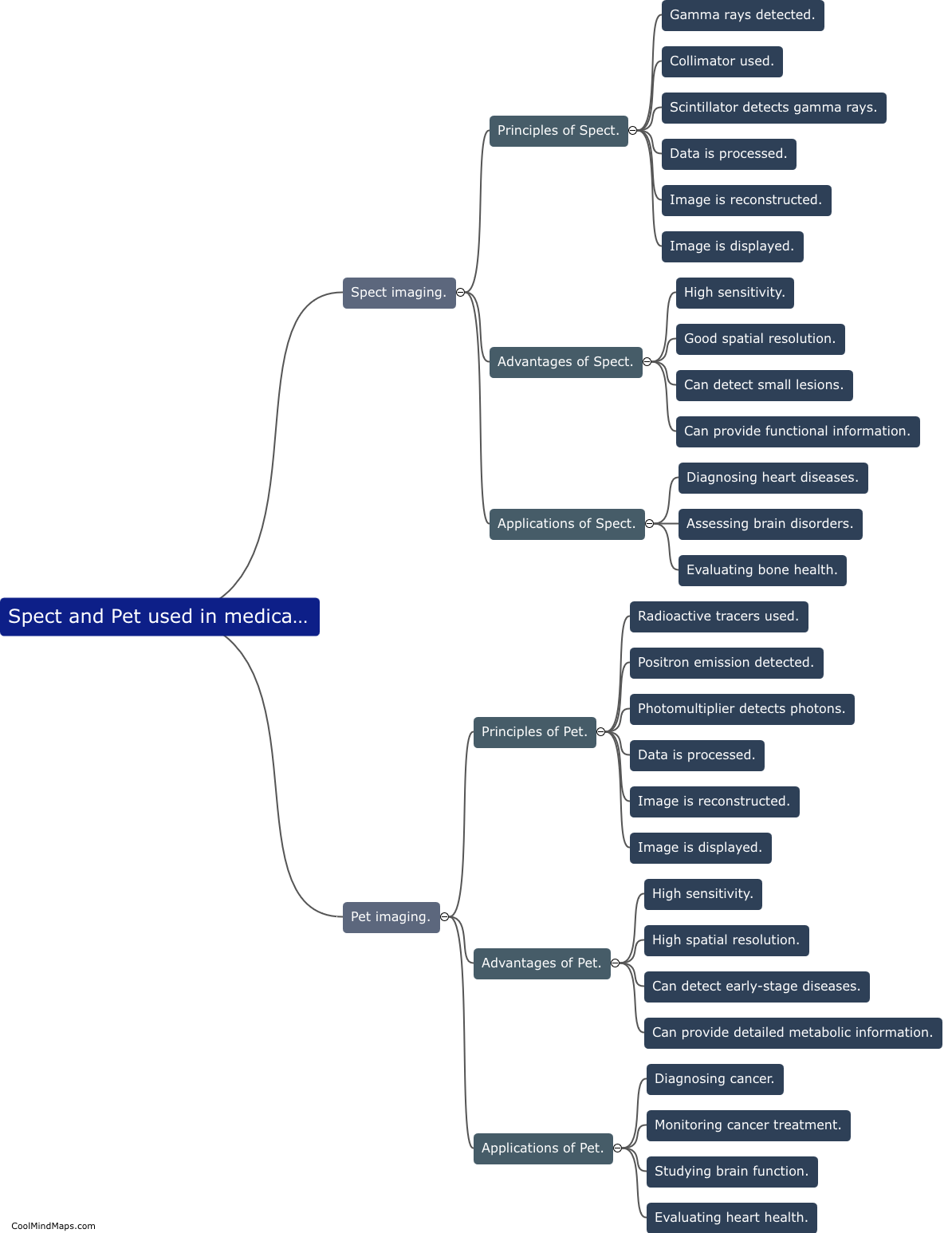
SPECT (single photon emission computed tomography) and PET (positron emission tomography) are two important techniques used in medical imaging for diagnostic purposes. SPECT involves the injection of a radioactive substance, which emits gamma rays. These gamma rays are detected by a specialized camera that rotates around the patient, capturing images from multiple angles. This enables the creation of three-dimensional images that provide information about blood flow, metabolism, and organ function. On the other hand, PET imaging utilizes a positron-emitting radioactive substance, which is injected into the patient's body. When the emitted positrons interact with electrons, they annihilate each other, releasing two photons. These photons are captured by detectors placed around the patient, allowing the creation of detailed images that highlight areas of abnormal cellular activity. Both SPECT and PET have proven invaluable in the diagnosis of various diseases, including cancer, cardiovascular disorders, and neurological conditions, as they provide clinicians with crucial information to guide patient management and treatment decisions.
This mind map was published on 12 November 2023 and has been viewed 93 times.
